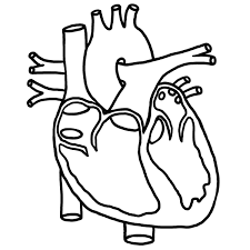
The Heart
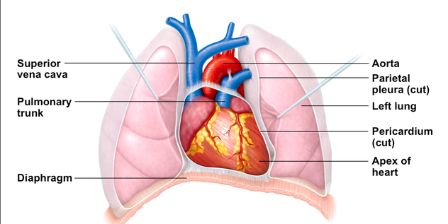
The heart is located in the thorax, between the lungs in the inferior mediastinum. It is approximately the size of a human fist. While its location is centered between the lungs, its orientation is not symmetrical. The apex is pointed down towards the left hip, which the base points upwards towards the right shoulder.
Coverings of the Heart
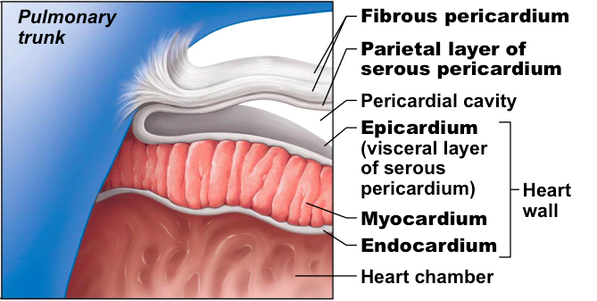
Pericardium - a double-walled sac
- Fibrous pericardium is loose and superficial
- Serous membrane is deep to the fibrous pericardium and composed of 2 layers
- parietal pericardium: outside layer that lines the inner surface of the fibrous pericardium
- visceral pericardium: next to the heart, also known as the epicardium
- Serous fluid fills the space between the fibrous pericardium and the serous membrane
Walls of the Heart
Three layers of the heart wall:
1. Epicardium
-outside layer
-this layer is the visceral pericardium
-connective tissue layer
2. Myocardium
-middle layer
-mostly cardiac muscle
3. Endocardium
-inner layer known as endothelium
1. Epicardium
-outside layer
-this layer is the visceral pericardium
-connective tissue layer
2. Myocardium
-middle layer
-mostly cardiac muscle
3. Endocardium
-inner layer known as endothelium
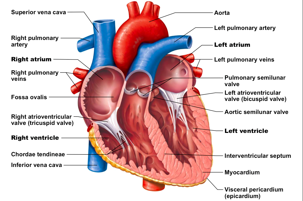
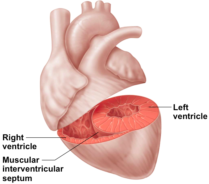
Chambers of of the Heart
The right and left sides of the heart act as separate pumps. There are four chambers in total: 2 atria and 2 ventricles. The atria (right & left) function as receiving chambers while the ventricles (right and left) function as pumping chambers.
|
The walls of the ventricles are more muscular than the atria because the ventricles must pump the blood and create enough force to move the blood where it needs to go. The left ventricle has a thicker muscular wall than the right ventricle because it pumps blood to the entire body, versus the right ventricle which pumps blood to the lungs. The interventricular septum separates the two ventricles. The interatrial septum separates the two atria.
|
test your knowledge of the anatomy of the heart here!
|
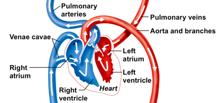
Great Vessels
Pulmonary trunk - blood is pumped out of the right side of the heart through the pulmonary trunk, which splits into right & left pulmonary arteries, carrying oxygen-poor blood to the lungs
Pulmonary veins - oxygen-rich blood returns to the heart from the lungs via the pulmonary veins
Aorta - oxygen-rich blood is pumped out of the heart to the rest of the body - the aorta has the highest blood pressure of all the vessels
Venae cavaea - oxygen-poor blood is returned to the heart through either the superior or inferior vena cava which both empty into the right atrium - these vessels have the lowest blood pressure of all of the vessels
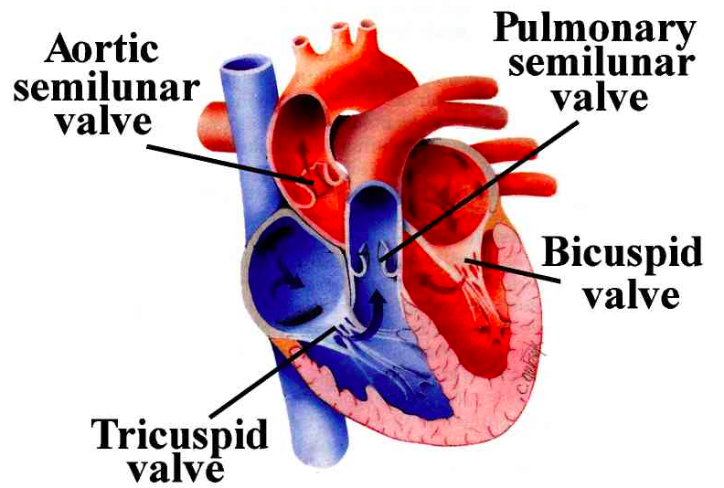
Heart Valves
|
Heart valves allow blood to flow in only one direction, to prevent back flow. There are 4 valves in the heart:
-Tricuspid valve - right side of heart
-Aortic semilunar valve (left ventricle) |
|
Cardiac Circulation
Blood inside the heart does not nourish the myocardium. The heart has its own circulatory system that brings it the oxygen & nutrients it needs to keep working. Cardiac circulation consists of:
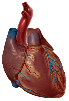
coronary arteries - branch off from the aorta to supply the heart muscle with oxygenated blood
cardiac veins - drain the myocardium of blood
coronary sinus - a large vein on the posterior of the hart, receives blood from the cardiac veins and empties oxygen-poor blood into the right atrium
Coronary heart disease happens when plaque builds up inside the coronary arteries, blocking blood supply to the heart muscles. This can cause angina and eventually a myocardio infarction (MI), commonly known as a "heart attack".
Blood Flow Through the Heart
- Blood enters the heart through the superior & inferior venue cavae into the right atrium.
- From the right atrium, through the tricuspid valve into the right ventricle
- From the right ventricle, blood leaves the heart as it passes through the pulmonary semilunar valve into the pulmonary trunk
- The pulmonary trunk splits into right & left pulmonary arteries, carrying (deoxygenated) blood to the lungs
- In the lungs, blood picks up the oxygen and drops off carbon dioxide
- oxygen-rich blood returns to the heart through the four pulmonary veins
- Blood enters the left atrium and travel through the bicuspid valve into the left ventricle
- From the left ventricle, blood leaves the heart via the aortic semilunar valve and aorta to travel to the rest of the body

|
How the Heart Actually Pumps Blood
|
Crash Course - The Heart, part 1
|
Crash Course - The Heart, part 2
|